Stories
Rita Allen Foundation Announces 2016 Scholars
Early-career biomedical researchers to examine the underpinnings of human behavior, cancer and chronic pain

The Rita Allen Foundation has named its 2016 class of Rita Allen Foundation Scholars, recognizing seven young leaders in biomedical science whose research holds great promise for revealing new pathways to advance human health. The selected Scholars will receive grants of up to $110,000 annually for a maximum of five years to pursue innovative research on the development and suppression of cancer, environmental influences on behavior, and the mechanisms and perception of pain.
These Scholars were selected for their original scientific approaches, focus on areas of global concern, opportunities for lasting outcomes, collaborations, and demonstration of leadership and learning potential.
Five of the Scholars were nominated by research institutions in the United States and selected by the Rita Allen Foundation’s Scientific Advisory Committee of leading scientists and clinicians. Two of the Scholars were selected for the Rita Allen Foundation Award in Pain in partnership with the American Pain Society. This marks the eighth year of this collaboration, which supports investigators who seek to understand and alleviate pain.
This year is also the 40th anniversary of the Scholars program, which began in 1976 as one of the first philanthropic fellowship programs of its kind for early-career biomedical researchers. The Rita Allen Foundation has provided funding to more than 150 young leaders in basic biomedical science, fostering creative research with above-average risk and promise. Scholars have gone on to make fundamental contributions to their fields of study, and have won recognition including the Nobel Prize in Physiology or Medicine, the National Medal of Science, the Wolf Prize in Medicine, the Lasker~Koshland Award for Special Achievement in Medical Science, and the Breakthrough Prize in Life Sciences.
“For 40 years, the Rita Allen Foundation has been investing in outstanding biomedical scientists at the early stages of their careers. With the class of 2016, we again are providing resources to these Scholars to pioneer bold approaches and innovative discoveries,” said Elizabeth Good Christopherson, President and Chief Executive Officer of the Rita Allen Foundation. “They will explore alternative treatments for childhood cancers, illuminate how diet changes our brains and affects our health, and investigate how chronic pain alters the central nervous system. These leaders join our community of Scholars, whose efforts to address basic biological questions are essential for improving health and reducing suffering caused by disease.”
The members of the 2016 class of Rita Allen Foundation Scholars are:
Steve Davidson, University of Cincinnati College of Medicine
Camila dos Santos, Cold Spring Harbor Laboratory
Monica Dus, University of Michigan
Katherine Hanlon, University of New England College of Osteopathic Medicine
Alex Kentsis, Memorial Sloan Kettering Cancer Center
Bo Li, University of North Carolina at Chapel Hill
Katharina Schlacher, The University of Texas MD Anderson Cancer Center
Meet the Scholars

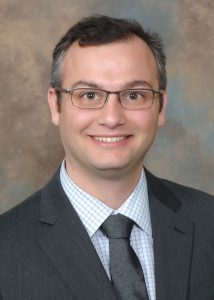
Steve Davidson
(Photo: University of Cincinnati)
Steve Davidson, University of Cincinnati College of Medicine
In conjunction with the American Pain Society
Assistant Professor of Anesthesiology
B.S., Psychology and Biology, University of New Orleans
Ph.D., Neuroscience, University of Minnesota
Project: How does the brain’s processing of emotion affect the perception of pain?
Steve Davidson dreamed of a career in ocean engineering until an undergraduate psychology course piqued his curiosity. He was intrigued by an image of a synapse—the intersection between two neurons. “Everything we do and think about and construct about our existence is due to these little chemicals that are flowing from one end of a neuron to another,” Davidson explains.
He signed up for more courses in psychology, biology and chemistry, and got involved in research on opiate dependence in rats and on motion sickness in humans. Davidson says he views pain as “philosophically interesting,” in that “it’s a fundamental experience but it’s hard to describe—it’s subjective.” He began to explore the emotional aspects of pain as a graduate student in Glenn Giesler’s laboratory at the University of Minnesota, where he examined how pain- and itch-sensing neurons in the spinal cord relay signals to the brain’s thalamus—including how scratching can suppress itch.
Now, Davidson seeks to reveal pathways to new therapies by applying knowledge about differences between the sensory and emotional dimensions of pain. Some patients with injuries to limbic areas of the brain, which surround the thalamus, experience pain asymbolia—a condition in which they are aware of pain but are not distressed by it. Davidson will study how neurons in the thalamus participate in the emotional perception of pain.
“Pain is a multitude of different things, and it may be possible for us to achieve better success if we pare down what we’re attacking in pain, and just focus on one aspect of it that causes the most suffering,” he says. “I believe that’s the emotional component of pain, and it may be possible to achieve this pain asymbolia effect in people through some therapeutic technique.”

Camila dos Santos
(Photo: Constance Brukin)
Camila dos Santos, Cold Spring Harbor Laboratory
Assistant Professor
B.S., Biology, Pontifical Catholic University of Campinas
Ph.D., Molecular and Cell Biology, University of Campinas
Project: What is the molecular basis of pregnancy-induced breast cancer protection?
As a graduate student in Brazil, Camila dos Santos pursued both master’s and doctoral research at the University of Campinas with hematologist Fernando Costa. In Costa’s lab, she honed in on the roles of genetic variation and gene expression in red blood cell development. As part of her graduate program, dos Santos visited Mitchell Weiss’ laboratory at the Children’s Hospital of Philadelphia, where she first observed the role of microRNAs during blood development. During a postdoctoral fellowship in Gregory Hannon’s lab at Cold Spring Harbor Laboratory, dos Santos sought to apply her knowledge of gene regulation and stem cell analysis to an underexplored area—breast biology.
“It was unbelievable to me that for such a relevant tissue, we knew very little about the different cell types in the breast, and there was very little we could do to really isolate those cells,” she recalls. She successfully established a technique to separate populations of breast cells, which enabled her to investigate gene expression during breast development, and especially during pregnancy.
Last year dos Santos set up her own lab at Cold Spring Harbor, where she and her team are continuing to study breast development in mouse models. They have found dramatic differences in the pace of breast development between first and second pregnancies, which appear to be mediated by epigenetic mechanisms—molecular changes that affect gene expression without altering DNA sequences. The Rita Allen Foundation award will allow the dos Santos lab to assess the relevance of these epigenetic phenomena for breast cancer risk, which is at least 30 percent lower in women who have a full-term pregnancy before the age of 25. A long-term goal is to use this knowledge to “propose a prophylactic strategy” so that even women who do not have an early pregnancy can “still leverage the preventive effect that the pregnancy signals are bringing to decrease the risk of breast cancer,” dos Santos says.

Monica Dus
(Photo: Pam Fisher/University of Michigan)
Monica Dus, University of Michigan
Monica Dus has been designated the Milton E. Cassel Scholar for the 2016 class of Rita Allen Foundation Scholars. This special award honors the memory of a long-time President of the Rita Allen Foundation who passed away in 2004.
Assistant Professor of Molecular, Cellular, and Developmental Biology
B.S., Biology, minors in Chemistry and Philosophy, University of Redlands Johnston Center for Integrative Studies
Ph.D., Biology, Watson School of Biological Sciences, Cold Spring Harbor Laboratory
Project: How does sugar in the diet influence feeding behavior?
Monica Dus got her first microscope at the age of 7, and got hooked on observing “the pervasive beauty of the natural world.” After a high school lesson on the lac operon, a trio of bacterial genes that regulates transport and metabolism of the sugar molecule lactose, “I started thinking about the continuous interaction between genes and environment, and how they shape everything,” Dus says.
She did Ph.D. research with Gregory Hannon at Cold Spring Harbor Laboratory, where she examined the roles of small RNAs in protecting animal genomes from transposons. These “jumping genes” can wreak havoc by disrupting vital genes and regulatory sequences. For her postdoctoral research, Dus decided to combine her interests in biology and philosophy by exploring the interplay among genes, environment and behavior.
“Eating is a quintessential example of a behavior that is changed by the environment,” she says. “And you can study it from a very simple and manageable perspective, because you can measure levels of nutrients and how they vary.” In Greg Suh’s lab at the New York University School of Medicine, Dus studied the neural circuits of hunger and satiety in fruit flies (Drosophila), and identified molecular mechanisms that allow animals to distinguish real sugar from fake sugar that does not provide nutrients.
Now, Dus and her group at the University of Michigan are using Drosophila as a model to understand how a high-sugar diet “deregulates the dynamic balance between hunger and satiety.” She hopes that her research into the neural, genetic and epigenetic regulation of feeding behavior will shed light on obesity and overeating—“not as a problem of willpower, but really as a problem of biochemistry—of sugar in the environment changing the brain persistently.”

Katherine Hanlon
(Photo: Ed Bilsky)
Katherine Hanlon, University of New England College of Osteopathic Medicine
In conjunction with the American Pain Society
Assistant Professor of Biomedical Sciences
B.S., Biochemistry and Molecular Biophysics, University of Arizona
Ph.D., Pharmacology, University of Arizona
Project: How do interactions between neurons and immune cells at the roots of spinal nerves impact sensitivity to pain?
Katherine Hanlon began her undergraduate studies as a business finance major, but an unfortunate turn of events inspired her to switch focus: her grandmother was diagnosed with cancer, and passed away just a few months later. “It completely changed the direction of my life,” Hanlon says.
She began working with pharmacology researcher Todd Vanderah, initially investigating the potential of compounds that activate opiate receptors to relieve cancer pain. This work “morphed into trying to figure out how the compounds we were using were actually stopping the growth of the cancer itself,” she says. It ultimately led Hanlon to focus on tumor immunology. She and her colleagues found that treating breast cancer cells with a drug that activates a protein called cannabinoid receptor 2 can cause the cells to commit cellular suicide.
In her own laboratory, Hanlon continues to examine how the activation of cannabinoid receptor 2 can curb the progression of cancer. With the Rita Allen Foundation Award in Pain, she and her research group plan to study the relationship between macrophages—immune cells that reside in all of the body’s tissues—and pain signaling in dorsal root ganglia, which are bundles of nerve cells found at the base of spinal nerves. They will evaluate how these macrophages may contribute to the development of chronic pain by triggering the recruitment of other immune cells to neural tissue.
“We’re looking at the native function of these tissue-resident macrophages, and we’re seeing that they do actually respond to a peripheral stimulus, like a peripheral injury,” Hanlon explains. “This is interesting, because most studies on the interaction between macrophages and the development of pain have looked at the site of injury, and not at the more central pathways themselves.”

Alex Kentsis
(Photo: MSKCC)
Alex Kentsis, Memorial Sloan Kettering Cancer Center
Assistant Professor
A.B., Biological Sciences, University of Chicago
S.M., Biochemistry, University of Chicago
Ph.D., Biophysics, New York University
M.D., Mount Sinai School of Medicine
Project: How does DNA transposition contribute to the development of childhood cancers?
Alex Kentsis is a physician-scientist who views the study of childhood cancers as a window into fundamental aspects of human development. “The beginning of my scientific career dealt with very basic scientific questions of self-organization of biological systems, and how biological macromolecules fold and attain their specific structures,” he says. As a doctoral student, he worked with structural biologist Katherine Borden to investigate the self-assembly of a class of proteins involved in leukemia and breast cancer.
Kentsis’ current research employs new technologies in genomics and proteomics to unravel cancer signaling pathways and explore therapeutic approaches to reengineer these pathways. His laboratory will use funding from the Rita Allen Foundation to examine the mechanisms and targets of a human DNA transposase. This enzyme enables the movement of transposons, or “jumping genes,” from one site in the genome to another, potentially altering the activities of genes that regulate growth and development. Kentsis and his colleagues recently discovered that this transposase exhibits abnormal activity in many embryonal tumors, which develop in the central nervous system in children and are thought to arise from residual embryonic cells.
“A specific conundrum in the field has been how childhood cancers, which are characterized by a paucity of gene mutations and occasional genomic rearrangements, can happen at such a young age, in cells that have not been exposed to many decades of environmental damage and errors in DNA replication,” Kentsis explains. His group’s findings on DNA transposase activity in these cancer cells “led to the hypothesis that perhaps such a gene is active in a subset of childhood tumors and induces these site-specific mutations.” A fuller understanding of the transposase may reveal novel oncogenes and tumor suppressor genes, which could point to new therapeutic strategies.

Bo Li
(Photo: Lars Sahl)
Bo Li, University of North Carolina at Chapel Hill
Assistant Professor of Chemistry
B.S., Biological Sciences, Beijing University
Ph.D., Biochemistry, University of Illinois at Urbana-Champaign
Project: Do molecules produced by gut bacteria modulate the human nervous system?
Bo Li has long been interested in antibiotic research as a fascinating convergence of chemistry and biology. As a Ph.D. student in Wilfred van der Donk’s laboratory, she focused on the enzymatic mechanisms that allow certain bacteria to produce lantibiotics (a class of peptide antibiotics)—in particular, nisin, which is widely used to prevent food spoilage. This experience “got me into studying fundamental science that has an impact on human health,” Li says. In her postdoctoral research with Christopher T. Walsh at Harvard Medical School, she delved into the genetics and biochemistry of another antibiotic, holomycin, in work that could inform strategies to produce antibiotics with novel mechanisms of action.
Now, Li and her colleagues are taking a multidisciplinary approach to discover new antibiotics and other bioactive molecules from a variety of sources. They conduct “genome mining” of microbial DNA sequences to identify biosynthetic pathways, and then use genetic engineering and chemical screening methods to characterize these new classes of molecules. The Rita Allen Foundation award will support a project to mine the genomes of bacteria that reside in the human digestive tract. These gut-associated bacteria “are already in the human body, so the small molecules they’re making will have an impact on human biology,” Li says. “But very little is known about this area. So my lab is building a program using genomics to identify specialized metabolites from these bacteria.”
Li’s group will search sequences from the Human Microbiome Project for genes involved in the production of molecules that may interact with receptors in the brain. One aspect of this work is aimed at finding molecules that target members of a large family of cell surface receptors, possibly leading to new treatments for mental and neurological disorders.

Katharina Schlacher
(Photo: MD Anderson Cancer Center/Medical Graphics and Photography)
Katharina Schlacher, The University of Texas MD Anderson Cancer Center
Assistant Professor of Cancer Biology
B.S., Microbiology, Karl-Franzens University
Ph.D., Molecular Biology, University of Southern California
Project: How do defects in the protection of stalled mitochondrial DNA replication forks impact the development of cancer and other diseases?
Early on in her biology training, Katharina Schlacher became captivated by DNA replication. “The entire process of adding nucleotides and having to copy by sequential addition was very interesting to me,” she says. “I thought it was such a fundamental process for life that it must be worth looking into more, even though a lot was already known about it.”
Schlacher first began to study DNA replication as a doctoral student in Myron Goodman’s laboratory, where she investigated E. coli genes involved in generating mutations induced by DNA damage. Around the time she completed her Ph.D., her mother was diagnosed with breast cancer. As Schlacher learned more about the disease, she realized that some tumor suppressor genes had similar functions to the genes she had studied in bacteria.
During a postdoctoral fellowship with Maria Jasin at Memorial Sloan Kettering Cancer Center, Schlacher discovered that the well-known breast cancer risk genes BRCA1 and BRCA2 were responsible for a previously unknown tumor suppression mechanism. This mechanism is separate from the genes’ functions in DNA repair, and involves the protection of stalled “replication forks”—dynamic sites where the two strands of DNA separate and are each copied through the action of specific enzymes. Protecting stalled replication forks from degradation prevents mutations and broader genome instability that can lead to cancer.
Since she established her own laboratory at MD Anderson in 2014, Schlacher has found evidence that defects in fork protection are present in breast cancers that lack classical BRCA mutations; these results could inform strategies to target cancers that are insensitive to current treatments. Funding from the Rita Allen Foundation will also allow Schlacher and her team to examine the importance of DNA replication fork protection in the mitochondria, the energy-producing compartments of cells. Schlacher has hypothesized that impaired fork protection in mitochondria may explain the increased risks for metabolic and cardiac complications observed in some breast cancer patients and those with Fanconi anemia, a blood cell production disorder linked to BRCA mutations.