Stories
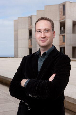
Axel Nimmerjahn – An Interdisciplinary Path to Make the Invisible Visible
They say a picture is worth a thousand words, but how do you create an image of real-time interactions inside the nervous system?
“For the brain and spinal cord, that’s a little challenging. Our goal is to shed light on this ‘black box.’ For a very long time, we simply didn’t have the technology [to study this], and so, we’ve developed that technology to open that box to look inside and see, how exactly does it work,” explains 2011 Rita Allen Scholar Axel Nimmerjahn.
Growing up in Germany, Nimmerjahn was exposed to medical science and technology at a very early age. His parents worked in a hospital and would share patient stories around the breakfast table and take him and his brother to conferences. “I think both of us liked this very much,” says Nimmerjahn. This science storytelling connected another passion, “one of the things that really inspired me as a teenager was a magazine called P.M. That magazine focused on communicating the latest findings in the natural sciences and technology to a broad audience. I really devoured those journals. They sparked my interest in the natural sciences and physics in particular.”
Nimmerjahn’s compulsory year of military service after high school opened opportunities to further explore his interests in science. He dedicated as much time as possible to attending medical science and physics lectures at local universities. “Education is essentially free [in Germany], so you can just walk into a lecture room and listen in. And so, that’s what I did,” he recalls. So captivated was Nimmerjahn by physics research that he declined an offer to attend one of Germany’s top medical schools in order to study physics—and never regretted it.
“At the same time, I never lost that interest in the medical sciences. So, after I graduated from university, I had the perfect opportunity to do both. I did both my master’s thesis, as well as my Ph.D., at the Max Planck Institute for Medical Research,” says Nimmerjahn. His training in physics complemented his interest in neuroscience as he began a journey into developing microscopes to image cells in the nervous system.
An accidental phone call to a Nobel laureate helped Nimmerjahn chart his path toward developing new technology to image and understand brain and spinal cord cells. Nimmerjahn was searching for a lab where he could complete his master’s thesis. Having heard of the Max Planck Institute’s research at the intersection of physics and biology, he called their department of cell physiology. “And the person who picked up the phone was Bert Sakmann. Dr. Sakmann is a Nobel laureate in physiology and medicine! Because his secretaries had already left for the day, he picked up the phone [when I called],” Nimmerjahn remembers.
Follow your passion. I think the Rita Allen Award in particular is one of those few opportunities where you’re encouraged to take risks, and you’re encouraged to follow your passion.
Sakmann became Nimmerjahn’s thesis advisor and introduced him to a new PI who was looking for students, Fritjof Helmchen. Helmchen had previously developed the first miniaturized two-photon microscope at Bell Labs and was looking to continue this work at the Max Planck Institute. With Nimmerjahn’s background in physics, he was an excellent fit for the project and was accepted into Helmchen’s lab as the first graduate student.
In addition to designing smaller microscopes, the Helmchen lab was working to develop complementary methods for labeling cells that would also allow researchers to view activity within cells. One method that was only beginning to emerge at the time was the use of viral vectors—a way to transfer genetic material into cells using the natural mechanism of how a virus infects a host cell. Nimmerjahn began a collaboration with another young investigator at the Max Planck Institute named Pavel Osten to use viral vectors to express fluorescent proteins in cells, such as the green fluorescent calcium indicator GCaMP, which changes its fluorescence intensity when bound to calcium ions, creating early images of activity within individual brain cells.
“We wanted to use the viral vector to express GCaMP1.6 in neurons. Once they have expressed it at high enough levels, we can mount the miniaturized microscopes [on the mice], and we can measure the [cellular] activity as the animal behaves. But these vectors have fairly broad tropism—they do not only infect neurons. They infect all sorts of other cells, including glial cells,” says Nimmerjahn. “One day, Pavel called me. He said, ‘Hey, you should come over. I have tissue slices where you can see the staining; it looks incredible.’ When I looked through the microscope, I said, ‘Yeah, you’re right, this is fantastic!’ The thing is, he was talking about neurons. I was looking at astrocytes, [a specialized type of glial cell] and no one at the institute at that time was interested in studying glial cells. I was looking at these astrocytes, these bushy cells that were sort of floating like clouds through the neurons’ dendritic trees, and that was my starting point into glial cells.”
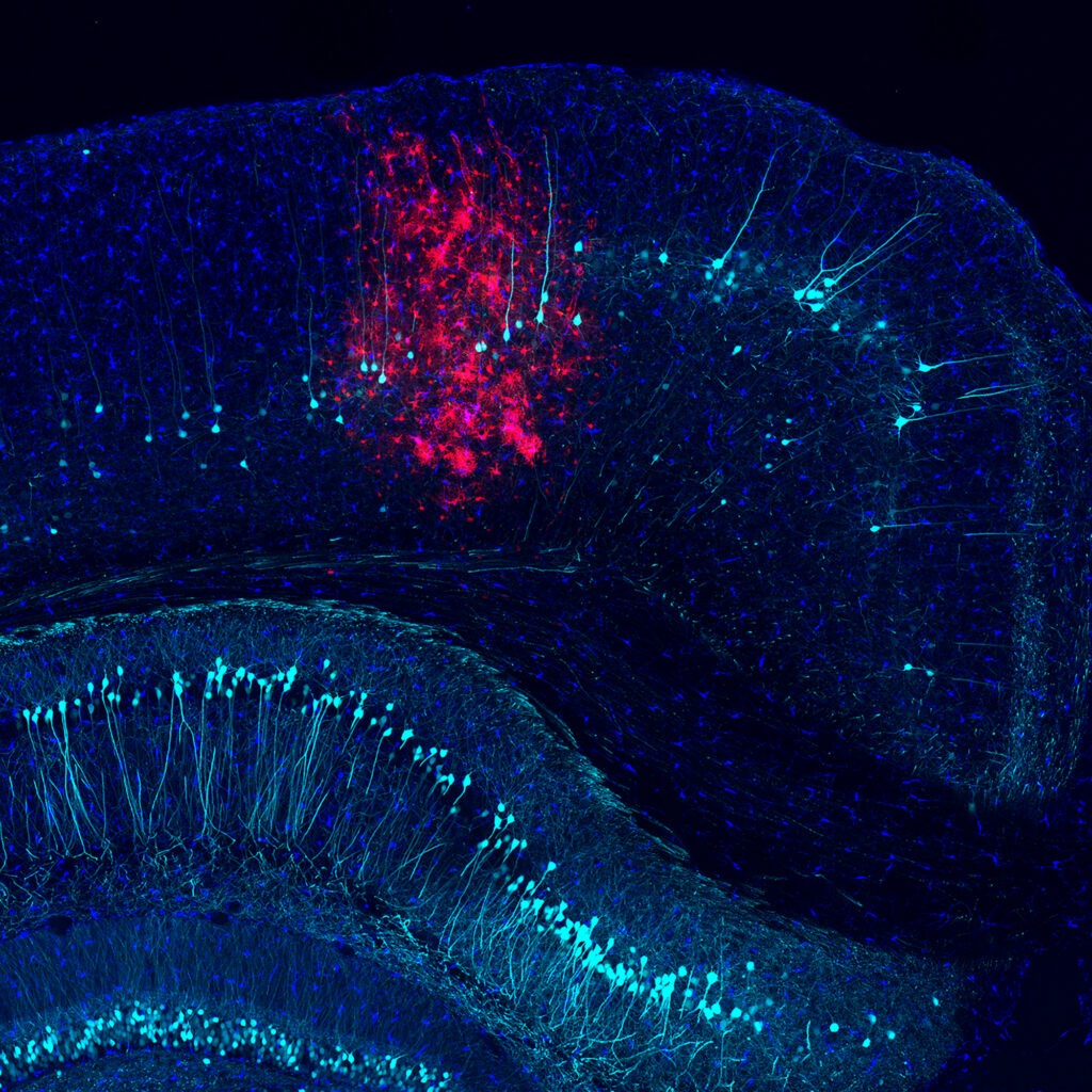
At the time, Nimmerjahn had limited prior knowledge of glial cells, support cells that interact with neurons in the central nervous system, and visited the library to learn more. He went on to make two important discoveries during his graduate work—identifying microglial cells as highly dynamic immune sensors within the brain and determining that certain synthetic dyes can be used to preferentially label living cell populations, including sulforhodamine 101, a dye to visualize astrocytes. “These two discoveries about microglia and then astrocytes turned out to be quite transformative for the field. We didn’t know at the time, of course. You only find out after many years what the impact of your research is, but those discoveries turned out to be quite impactful,” remarks Nimmerjahn.
Nimmerjahn went on to complete a postdoctoral fellowship at Stanford University in biology and applied physics, furthering his interest in working at the intersection of these two fields. In 2010, he joined the Salk Institute to start his own lab and in 2011, he was selected as a Rita Allen Scholar. Reflecting on the process of becoming a Rita Allen Scholar and attending his first Scholars Symposium as part of the community, he shared, “It was just a fantastic meeting. It really felt like a family, and that’s very difficult to achieve. I still have contact with people from that meeting, and some of them even invited me to give a talk. One of the participants, Greg Lemke, is an investigator here at Salk and a former Rita Allen Scholar. We ended up collaborating and we have published three papers that further studied microglia and the mechanisms that underly their immune surveillance. There’s many, many connections beyond the actual funding period where the Rita Allen community really had a big impact on my career.”
Nimmerjahn is now an associate professor in the Salk’s Waitt Advanced Biophotonics Center. When he first arrived at the Salk Institute, he decided to extend his research from the brain into the spinal cord. “The spinal cord is a black box, right now. It’s very difficult to record from the structure because it moves quite a bit. Electrophysiology requires that you stick an electrode into the tissue, but if the tissue moves a lot, that’s a problem. We’ve developed three generations of wearable microscopes. In 2016, we published the very first paper that showed that it’s possible to record cellular activity in the spinal cord of a behaving animal. We showed that you can watch in real-time how cells in the spinal cord encode sensory information,” recalls Nimmerjahn.
One discovery in the spinal cord showed that astrocytes display unexpected forms of excitations or stimulus responses. With an innocuous sensory stimulus, astrocytes responded predictably in a way that mirrored surrounding neurons. “The biggest surprise was when we then kept increasing the stimulus intensity, it transitioned from innocuous to becoming noxious or painful; then the astrocyte response is very different. You see an excitation that you wouldn’t see in the local neurons. It was a really widespread excitation, and we now know that that excitation plays a role in regulating pain sensation.”

In a paper on bioRxiv as a pre-print, Nimmerjahn and colleagues demonstrate that it is possible to decode what an animal did based on the information it processed on a behavioral task with 80 to 90 percent accuracy just by looking at astrocytic calcium excitation, an ion concentration change that can be captured through imaging technologies, in the motor cortex. Something similar may be the case for the spinal cord, where astrocytes show region-specific excitation to different types of sensory and motor information. Reducing this excitation in spinal sensory areas by inhibiting specific receptor pathways alleviates pain, says Nimmerjahn. “Once you know the underlying mechanisms, then you can go in and either reduce pain or prevent the transition from acute to chronic pain, or if chronic pain has already developed, revert it. So, that’s one of the main goals of our current studies.”
Here Nimmerjahn shares his advice to young researchers, what he enjoys doing outside of the lab, and insights on the impact of the Rita Allen award.
What advice would you give to other early career scientists thinking of applying for the Rita Allen Award?
Follow your passion. I think the Rita Allen Award in particular is one of those few opportunities where you’re encouraged to take risks, and you’re encouraged to follow your passion.
Are there any interests or things that you like to do when you’re not in the lab that you would like to share?
One of the perks that comes with this job is travel. [When you attend an international conference] you get the additional perk of having a local that can introduce you to the culture—and that means not only the food, but also the way the society works. That’s something that I really thank my parents for, as well. They took us on all sorts of trips when we were really young, and so, early on, you get exposed to all these different cultures. It really sort of makes you think about the things that you’re taught when growing up—is this the only way, or is this even the right way of doing things?
How would you say the award has influenced your career trajectory as well as your research?
Having that stamp of approval, early on helps. So, following the Rita Allen Award, I received, in 2012, a New Innovator Award from NIH. Then, the following year, an Exceptional, Unconventional Research Enabling Knowledge Acceleration Award—this is an appreciation that stands for exceptional research. And now, fast forwarding, the most recent award I received as a team director, is a U19 Award from the NIH. It’s an $11.2 million grant, which involves four different investigators [including another Rita Allen Scholar, Lin Tian (2014)]; it’s team-based research involving a protein engineer, a computational scientist and theorist, and molecular biology and genomics researcher. We hope that [our project] has a big impact on the field because we now have a protected space to do something that an individual lab can’t do.